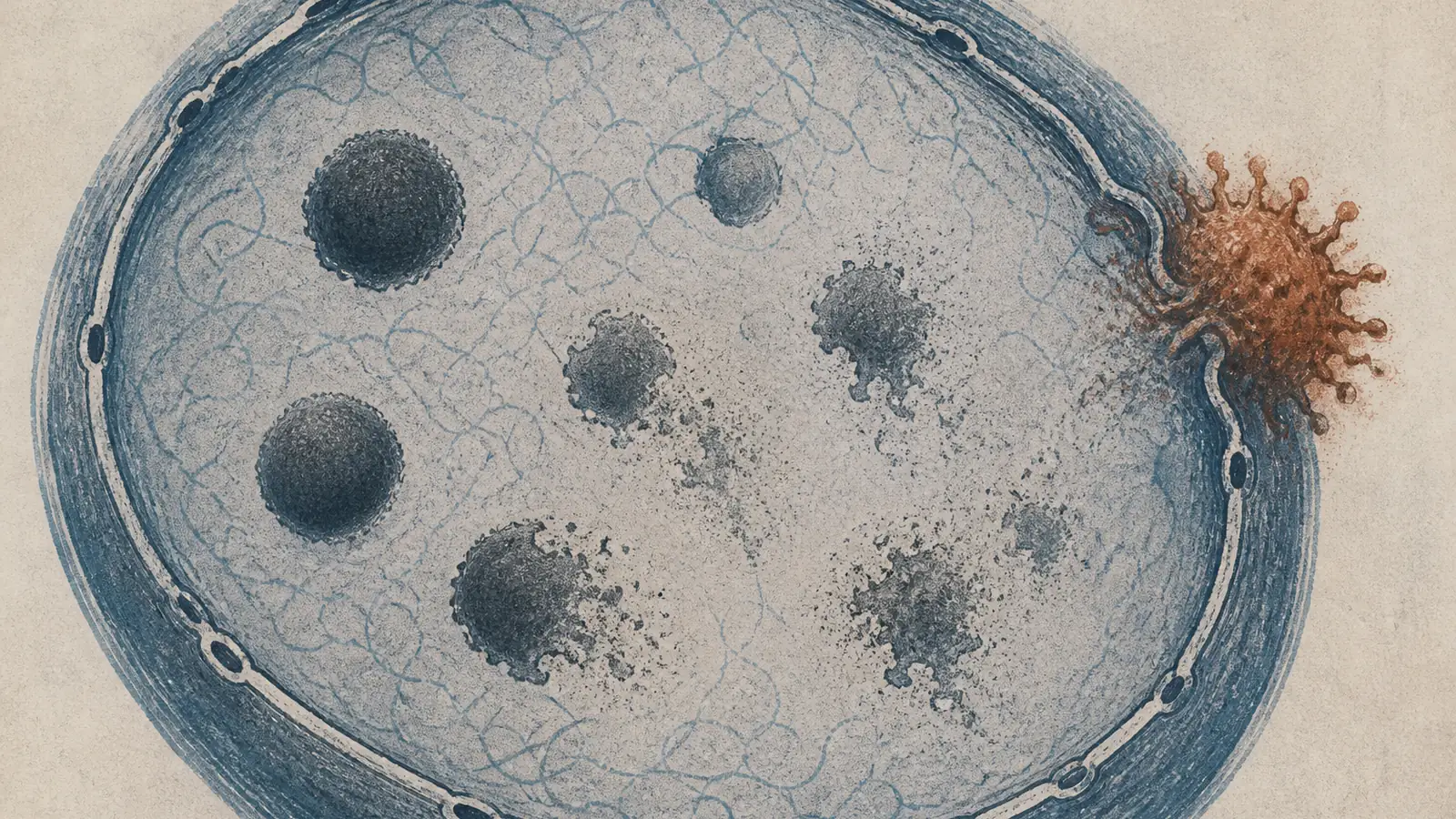
Iuliia Kotova watched the little droplets in the nucleus dissolve and disappear, then watched it happen again, in every cell line she tried and with every strain of influenza she threw at them. The structures are called paraspeckles, and the vanishing act was so consistent that it stopped looking like collateral damage and started looking like a plan.
The method is new. The drugs are not.
“What surprised us most was the paraspeckles,” Kotova, the first author and now at ETH Zurich, told EMBL. “Watching these tiny organelles in the nucleus dissolve, consistently across every cell line and every flu strain we tested, told us this isn’t a side effect of infection – it might be a strategy.”
Side effect versus strategy is the distinction the whole paper turns on. A virus breaks a lot of things on its way through a cell, and most of the breakage is incidental, the mess of a hijacking rather than the point of it. Kotova’s group, based at EMBL Hamburg and working with the Leibniz Research Institute for Molecular Pharmacology in Berlin, argue in Nature Microbiology that this particular demolition is too consistent to be an accident. Something about influenza appears to be actively driving it.
Paraspeckles are worth understanding before deciding how impressed to be. They are membraneless compartments, droplets that condense inside the nucleus around a long strand of non-coding RNA, and their day job is partly janitorial: they soak up RNA-binding proteins and hold them out of circulation. Some evidence suggests the same droplets help tune the cell’s stress and antiviral responses. So watch what dismantling them would buy the virus. The RNA-binding proteins spill back out, and, as the EMBL team describes it, the virus can then put them to work replicating its own genome. Group leader Jan Kosinski adds that “there may also be a second benefit for the virus”: if the paraspeckles were part of the cell’s alarm system, tearing them down would silence the alarm in the same motion. Free the labor, kill the warning, one move. If that reading holds, it is an unusually tidy piece of sabotage.
The reason the team could see any of this is the part virologists will envy. For years the standard way to catalog which viral proteins grab which human proteins has been to break the cells open and fish the pairs out of the resulting soup. The trouble with the soup is obvious once you say it out loud: proteins that never meet inside a living cell land in the same tube and can grab each other anyway, producing tidy interaction maps of encounters that never happen. Kotova’s collaborators Boris Bogdanow and Fan Liu built a version of cross-linking mass spectrometry that chemically locks protein partners together while they are still inside intact, infected cells, before anything is broken open. “XL-MS allows us to capture protein-protein interactions directly in infected intact cells, while also providing structural information,” Bogdanow said. They then fed those real contact points into a modified version of AlphaFold, so the structure-prediction software was working from data about where proteins actually touch instead of guessing. Influenza replication is thought to run on interactions between up to fourteen viral proteins and their human partners; this is a map of them drawn from inside the room instead of from the hallway.
The nucleus was not the only place it paid off. The map also traced how hemagglutinin, the virus’s surface protein, gets shepherded and chemically finished by the host machinery of the endoplasmic reticulum and the Golgi, the cell’s protein-processing line. Hemagglutinin is the spike the immune system learns to recognize and the target most flu vaccines are built around, so knowing which host factors it leans on to mature correctly is not a footnote.
This is where a veteran reader should slow the enthusiasm, because the press materials do not. The releases reach fast for “new drug targets” and for the specter of “dangerous flu strains,” and both are carrying more than the data can bear. This was a lab-adapted strain of influenza A in cultured human cells: no antiviral was tested, no target validated as druggable, no animal exposed to it. What the team produced is a high-resolution atlas of interactions and one strong hypothesis about paraspeckles, which is a contribution, and an honest one. The method is new. The drugs are not.
The scientists are careful about this in a way the coverage is not. Bogdanow’s own framing is that the study “lays the groundwork to apply the methodology to viruses of potential pandemic relevance, such as H5N1”, and groundwork is the right word. H5N1, the bird-flu strain that has spent months moving through American dairy herds, was not part of this study; it is where the authors would like the method to go, not a place it has been. His other claim on the work’s behalf is just as hedged: the map, he says, may “through structural modelling, help identify actionable targets for future pharmaceutical interventions.” May. Help. Future. Those are the load-bearing words, and they belong to the scientist, not to the headline.
One detail reads well against the usual backdrop of this beat. The public writeups foreground academic and public institutions rather than a pharmaceutical sponsor: EMBL, a Leibniz institute, the Charité, ETH Zurich, chasing a basic question about how a virus rewires a cell. Whatever the funding lines turn out to say, that is how a drug target is supposed to be found in the first place, years before anyone has a molecule to sell, in a lab where the surprise on the screen is allowed to be the point.
Kotova has since moved to ETH Zurich. The map she helped draw sits where useful maps sit at this stage, describing the territory in more detail than anyone had before and promising nothing about the road. What it holds, that the field did not have a year ago, is a single reproducible image: the droplets in the nucleus, dissolving on cue, every time. Whether anyone turns that into a drug is the next decade’s problem, not this paper’s.
Sources
- Nature Microbiology – Kotova et al., “Mapping in-cell protein contact sites reveals hijacking of paraspeckles during influenza A virus infection” (2026)
- EMBL – “Scientists map how the flu virus rewires the human cell from the inside” (2026)
- ScienceDaily – “New molecular map reveals how the flu virus hijacks human cells” (2026)
- Genetic Engineering & Biotechnology News – “Flu Virus Interaction with Host Cell Machinery Mapped Inside Infected Cells” (2026)
- EurekAlert! – “Scientists map how the flu virus rewires the human cell from the inside” (2026)
- Nature – “Bird flu virus has been spreading among US cows for months, RNA reveals” (2024)