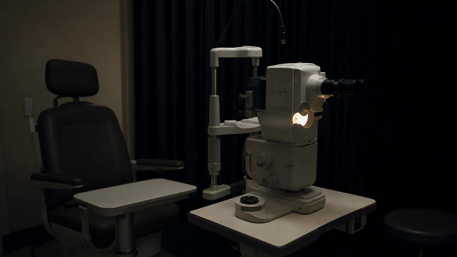
Sit an otherwise healthy 68-year-old in a dark room, shine a bright light to bleach out the retina, and then time how long the eye takes to find its way back to seeing in the dark. Most people recover in about 9 minutes. Some take 15. The ones who take 15 look, on every scan you can run that day, entirely normal. Their maculas are clean, their vision is fine, and they are roughly twice as likely to have early macular degeneration three years later as the people who recovered fast.
That is not a wellness slogan. It is the Alabama Study on Early Age-Related Macular Degeneration, 325 adults aged 60 to 86, followed for three years, funded by the National Eye Institute and the National Institute on Aging and published in 2015. The researchers drew a line at a rod-intercept of 12.3 minutes: slower than that counted as abnormal dark adaptation. People on the wrong side of the line developed early AMD at nearly double the rate, a relative risk of 1.99, with a confidence interval that stayed above 1.0 even after the authors adjusted for age and smoking. A 2025 follow-up found the same thing and added a detail that ought to make clinicians uncomfortable: of every visual function tested, rod-mediated dark adaptation was the only one that tracked both the onset and the early progression of the disease in healthy-looking eyes.
The scan was clean. The rods were not.
Here is the part that should bother anyone paying for eye care. The test is cheap, it costs a dark room and about ten minutes, and the public paid for the science behind it more than a decade ago. It is still not part of a routine eye exam. When a validated, inexpensive, publicly funded early-warning test sits unused that long, someone eventually packages it, names it, and adds a blood draw. That someone, this month, is an optometrist in Utah.
Walker L. Shaffer has been publishing a column series in the optometry trade press building out something he calls “retinal reserve,” and his July 7 installment lays out the operational version. He defines retinal reserve as the retina’s capacity to keep functioning despite the cumulative burden of metabolic, inflammatory, vascular, and oxidative stress. His organizing claim is the one the Alabama data support cleanly: functional decline often precedes visible structural damage, so by the time a lesion shows up on imaging, the window has already closed. He proposes catching it earlier with a battery of functional tests, contrast sensitivity, dark adaptation, electroretinography, and then reaching for the bloodwork.
The bloodwork is where the framework gets ambitious. Shaffer’s core panel, ordered on essentially every patient, runs HbA1c, fasting glucose, a lipid panel, homocysteine, hs-CRP, erythrocyte sedimentation rate, and an omega-3 index, with disease-specific add-ons layered on top: kidney markers and apolipoprotein A1 for macular degeneration, morning cortisol and advanced glycation end products for glaucoma suspects. It is a coherent idea. It is also, for now, only an idea, published as a practitioner’s column rather than a validated protocol, with no defined threshold for what a “low reserve” score would be and no trial testing whether ordering that panel changes what happens to anyone’s vision.
The instinct behind it is not wrong, and it is worth being fair about that. The eye is the one place in the body where a clinician can look at central-nervous-system tissue and living blood vessels directly, without cutting anyone open, and the research establishment has spent the last few years confirming how much that view reveals. Functional retinal changes are being studied as potential early markers of Parkinson’s disease, turning up in the retina’s electrical signaling and proposed as a signal that could precede the motor symptoms that define the diagnosis. A 2025 review in Alzheimer’s & Dementia catalogues structural, functional, and molecular retinal changes tied to cognitive decline. The retinal microvasculature has been linked to tubular markers of kidney injury.
So the systemic framing is not the weak part. The idea that homocysteine and chronic inflammation and omega-3 status are doing quiet damage years before anyone gets a diagnosis is the same thinking that mainstream ophthalmology has been slow to carry into an exam room, and Shaffer is applying it to an organ where the damage is unusually easy to see. The problem is narrower and more specific than the frame.
The gap here is not between good science and quackery. It is between a validated functional test that almost no one runs and a broad commercial panel that no one has validated. Dark adaptation earned its place in the literature over a decade of federally funded prospective follow-up. The seven-analyte “reserve” workup bolted alongside it has earned exactly none of that yet, and the difference matters, because a framework that folds one rigorously tested measurement into six untested ones tends to launder the credibility of the first onto the rest. A patient who hears “retinal reserve panel” has no way to know which number in it actually predicts anything, and right now only one of them does.
That is the real trouble with the panel: it exists to fill a vacuum that never should have opened. The dark-adaptation test works, it is cheap, the public already bought it, and a decade on it still hasn’t reached the patients it was built for. Shaffer is early and he is ahead of his evidence. He is also asking the question the field left lying on the table: if the retina starts failing before the scan can see it, why is no one looking until the scan does?
Sources
- Healio – Shaffer, “Measuring retinal reserve: practical framework for functional testing, systemic biomarkers” (July 7, 2026)
- Ophthalmology / PMC – Delayed rod-mediated dark adaptation as a functional biomarker for incident early AMD, ALSTAR cohort (2015)
- Ophthalmology – Delayed dark adaptation associated with AMD incidence and early progression, ALSTAR2 (2025)
- Neurobiology of Disease – Early detection of Parkinson’s disease: retinal functional impairments as potential biomarkers (2025)
- Alzheimer’s & Dementia – Retinal biomarkers in cognitive impairment and dementia: structural, functional, and molecular insights (2025)
- Kidney360 – Tubular biomarkers and retinal microvasculature (2025)
- Frontiers in Ophthalmology – Editorial: Retinal biomarkers of neurodegenerative diseases (2024)