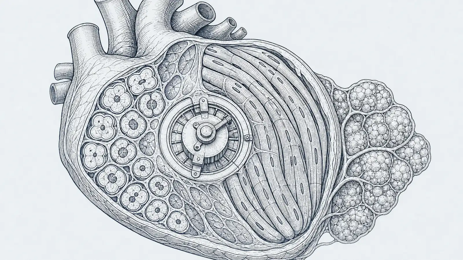
I used to file “your heart can’t heal itself” next to “teeth don’t grow back,” in the drawer marked permanent. You have a heart attack, the starved muscle dies, and instead of new muscle you get scar. Zebrafish regrow heart tissue. Newborn mice can do it too, for about a week before the window closes. Adult humans just patch the hole with collagen and live with a weaker pump. That always read to me like a wall.
It is not a wall. It is a tradeoff, and a team in Japan just found the knob that sets it. Stranger still, the knob turned out to be a gene I had only ever met in the world of body fat.
Here is what I did not appreciate until I sat with the biology. A heart cell cannot be good at two jobs at once. It can keep its machinery loose enough to divide, or it can pour its resources into the dense sarcomeres and packed mitochondria it needs to contract hard for eighty years. Maturing and multiplying pull against each other. The reason your adult heart does not regenerate is that its cells grew up, and growing up meant quitting the cell cycle. So the real question was never why heart cells can’t divide. It was what tells them to stop.
A group led by Kanae Tani, with Yoshinori Yoshida at Kyoto University’s iPS cell institute and Antonio Lucena-Cacace in Osaka, went looking for that signal. Writing in Stem Cell Reports, they grew human heart cells from iPSCs, wired them with fluorescent reporters that light up depending on where a cell sits in its division cycle, and then did the simplest, most revealing thing you can do. They turned one gene up, turned it down, and watched.
The gene is PRDM16. Turn it down and the cells drift back toward dividing: proliferation regulators like CDK1 and phospho-AKT climb, and the cells regain the competence to multiply. But those same cells build sloppy sarcomeres, their mitochondria falter, and in engineered heart tissue they squeeze weakly. Youthful and dividing, not much good as muscle. Turn PRDM16 up and it flips: proliferation shuts down, the cells grow larger, they switch on the adult contractile protein TNNI3, ramp up oxidative metabolism, and settle their twitchy spontaneous beating toward a calmer adult rhythm. Mature, strong, and done dividing. One transcription factor sitting on the exact fulcrum between the two states, working like a rheostat instead of an on/off switch.
And then the name stopped me, because I already knew it from somewhere completely different. PRDM16 is the master switch for brown fat: drop it into the right precursor cell and you steer it toward becoming a calorie-burning, mitochondria-stuffed brown fat cell instead of muscle. So why would one zinc-finger protein be the maturity referee in two tissues as unalike as fat and heart? The more I chewed on it, the less coincidental it looked. Both jobs are the same kind of decision: a cell committing to a high-energy, mitochondria-dense adult identity and giving up its other options to get there. PRDM16 may just be what that commitment looks like at the level of DNA, wherever a cell has to choose between staying flexible and getting serious about metabolism.
This is not abstract. Losing PRDM16 already breaks human hearts. Mutations in it drive the cardiomyopathy in 1p36 deletion syndrome, the most common terminal chromosome deletion at roughly one in 5,000 births, and between 23 and 27 percent of those children develop cardiomyopathy. The same gene turns up mutated in nonsyndromic left ventricular non-compaction and dilated cardiomyopathy too. So the dial these researchers were turning in a dish is the same one that, stuck in the wrong position in a real child, produces a failing heart.
of 1p36 deletion children
- Lower bound
- 23
- Upper bound
- 27
I want to keep my enthusiasm honest, though. This is human heart cells in a culture well and thumbnail-sized engineered tissue, not a patient, not even an animal. A rheostat that behaves beautifully in a dish is a long way from anything you could give a person after a heart attack. And the tradeoff itself is the trap: you cannot just crank PRDM16 down to spark division, because low PRDM16 also means weaker muscle. Any real therapy would have to divide the cells first and then re-mature them, a question of timing and sequence, not just which way you push. Nobody has shown that.
One thing I always check, and it cuts the friendly way here: who paid. Not a drug company with a candidate to sell. The Leducq Foundation and Japanese public science agencies funded this, the kind of basic-mechanism work done to understand something rather than move a product. That is part of why the paper calls PRDM16 an interesting position on a continuum rather than a cure, and I find that restraint more persuasive than any splashy claim.
So no, I am not going to tell you your heart can regrow. But I have moved “adult hearts can’t heal” out of the permanent drawer and into the one labeled “we now know the reason, and reasons can be worked on,” which is a genuine shift for me. And here is the one piece already worth acting on: if someone in my family carried a PRDM16 variant, I would push for cardiac screening now, not because a dish experiment says so, but because the clinical genetics already does. The rest, I will believe when it works in an animal.
Sources
- Stem Cell Reports – Tani et al., PRDM16 modulates aspects of cell cycle dynamics and maturation in human iPSC-derived cardiomyocytes (2026)
- American Journal of Human Genetics – Arndt et al., Fine mapping of the 1p36 deletion syndrome identifies mutation of PRDM16 as a cause of cardiomyopathy (2013)
- Nature – Seale et al., PRDM16 controls a brown fat/skeletal muscle switch (2008)
- Science – Porrello et al., Transient regenerative potential of the neonatal mouse heart (2011)
- 1p36 Deletion Syndrome and Left Ventricular Non-compaction Cardiomyopathy: two case reports (2021)
- News-Medical – PRDM16 regulates the balance between proliferation and maturation in human cardiomyocytes (2026)