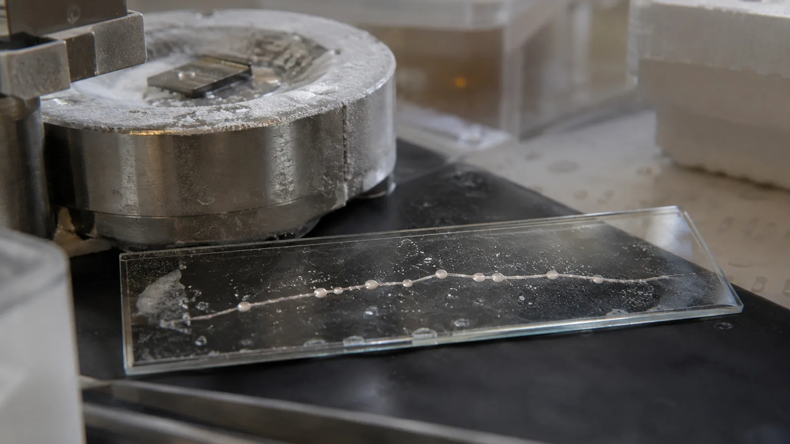
I have been carrying around a wrong picture of my own brain for most of my life, and so, almost certainly, have you. It is the picture in every textbook: the neuron as a little octopus, a round body with long smooth cables trailing off it, each one a clean tube carrying an electrical pulse from here to there. I drew that tube in a college notebook. My professors drew it. Santiago Ramón y Cajal, the Spanish anatomist who basically founded the field, drew a version of it more than a century ago. And it turns out the tube might be the part we got wrong.
A team at Johns Hopkins, led by cell biologist Shigeki Watanabe with graduate student Jacqueline Griswold as first author, reported in Nature Neuroscience that the unmyelinated axons of the mammalian brain, the bare fibers with no insulating sheath, are not smooth at all. Across the stretches they imaged, the axon bulges into a repeating series of tiny swellings connected by a thin cable, like beads knotted onto a string. The full paper puts numbers on the beads: each swelling runs about 200 nanometers across, and the connecting thread between them narrows to roughly 60. On the scale of a cell that is not a rounding error. It is a fundamentally different shape.
Here is what makes that unsettling rather than merely tidy. Bead-like swellings along an axon already have a meaning in neuroscience, and it is a grim one. Injured and dying neurons “bead up,” and for decades those bumps have been read as a signature of damage. If the beads are the normal resting shape, then some of what we have been calling injury in images of aging and diseased brains may be ordinary anatomy nobody knew to expect.
So my first question, the one I could not let go of, was how we missed this for a hundred years. The answer is almost embarrassingly physical. To photograph a structure a hundred times thinner than a human hair, you have to fix the tissue first, and standard chemical fixation slowly pulls water out of the cell. A dehydrated bead flattens toward a smooth tube. Watanabe’s team instead froze the tissue cold in milliseconds under high pressure, catching the structure where it actually sits. When they went back and used the ordinary imaging chemical, the pearls vanished entirely, which may be exactly why nobody had seen them. They found the same beaded pattern in mouse neurons grown in a dish and in brain slices from mice of various ages.
Here is where the biology stopped being tidy and got genuinely strange. Working with theoretical biophysicist Padmini Rangamani at UC San Diego, the team built a mathematical model of the axon membrane and asked what shape it wants to take on its own. The answer was: this one. In their model nothing had to hold the beads in place from the inside, no protein skeleton, no scaffold. Just the physics of a thin oily membrane under tension, the same force that makes a stream of water off a tap break into droplets, pulling the tube into pearls. Wait, so the shape of the wire your thoughts travel down is set by surface tension rather than by machinery? That is the part that got me, and the model earned it: when they stripped cholesterol out of the membrane and changed its stiffness, the pearling smoothed out and the axon’s ability to carry a signal dropped with it. The shape was not decoration. It was doing something.
And it moves. When the researchers drove the neurons hard with high-frequency electrical bursts, the pearls did not sit still. They swelled, about 8 percent longer and 17 percent wider, and stayed puffed up for at least 30 minutes after the stimulation stopped. The signal running down the line sped up as they swelled. So the geometry is not fixed plumbing you are issued at birth. It flexes with how hard the line has been worked, and that flex tunes how the signal runs. A wider bead gives ions more room to move; pinch the cable and you change the traffic. Your axons remodel their own shape around your activity, on a timescale of minutes.
Not everyone is sold, and the objection is exactly the one you would want raised. Bead-like swelling is the classic sign of a stressed or injured neuron, so critics have asked whether the pearls are real resting structure or an artifact of culturing and imaging, which is why some outlets have flagged the work as controversial. The team’s answer is the strongest thing they have: they imaged live cells that were never frozen and never chemically fixed, and those showed the pearls too. If the beads survive when you touch the cell as little as physically possible, “artifact of the preparation” gets hard to sustain. The tension has not fully resolved. It is the live edge of the debate, not a footnote.
But sit with what it would mean if the pearls are ordinary. For a century, part of the “damage” we read in images of aging and diseased brains may have been normal structure, and the actual disease signal would be a change in the beads rather than their mere presence. The map of how signals travel through unmyelinated fibers, the ones that make up a huge fraction of the brain and were always treated as the plain, boring wiring, would need redrawing. The work ran on public research money, the NIH among the funders, basic science with no drug at the end of it, which is exactly the kind of quiet, foundational looking that turns out to matter most.
I came into this ready to file it under “cool microscopy trick, textbook gets a tweak.” I am leaving convinced it is bigger, and here is my ownable position: the next time I see a brain scan described as showing “axonal beading” as proof of injury, I am going to ask which beads, and compared to what. The plain tube I memorized was a shortcut nobody meant to lie with. But it was still wrong, and I would rather carry the messier, truer picture in my head, pearls and all.
Sources
- Nature Neuroscience – Griswold et al., “Membrane mechanics dictate axonal pearls-on-a-string morphology and function” (2024)
- PMC – full text, Griswold et al. (pearl and cable dimensions)
- Johns Hopkins Medicine – study announcement (high-pressure freezing method)
- Neuroscience News – cholesterol, membrane mechanics, activity-induced swelling, and conduction
- ScienceAlert – the artifact-vs-real-structure debate and the live-cell rebuttal
- bioRxiv – preprint of the axon-pearling work
- ScienceDaily – coverage and funding note