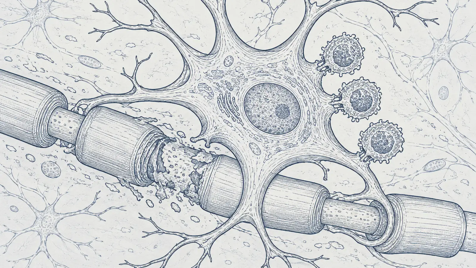
For most of my life I filed astrocytes under packing material. The name sounds decorative on purpose: astro, star, the little star-shaped cells that supposedly just hold neurons in place, mop up spills, and keep the metabolic lights on. Support staff. Scaffolding. The stuff between the important parts. So when I learned that inside a multiple sclerosis lesion these same cells can dress up like professional immune cells and help hand the nervous system to the T cells attacking it, I had to sit with how wrong the cartoon was.
The tell was in a title. A late-September research highlight in Nature Immunology called Presenting astrocytes, and “presenting” there is not a flourish. It is the technical verb. Antigen presentation is the immune system’s way of saying “here, look at this, attack it.” These cells do that.
The study it crowned lands the point hard. In work published in Nature in August, a team led by Joon-Hyuk Lee, Francisco Quintana and colleagues reported that a specific population of astrocytes, the ones flying both CD40 and MHC-II on their surface, actively push the brain’s immune system to turn on the brain. Using rabies-based barcoding to catch cells in the act of touching, single-cell sequencing, and genetic knockouts, they showed astrocytes talking directly to CD4+ T cells and sharpening the pathogenic Th17 response that does the damage in experimental autoimmune encephalomyelitis, the standard mouse model of MS. Switch those astrocytes off genetically and the disease eases.
Then came the part I did not see coming, and it runs on metabolism, which is the last place I expected a story about the immune brain to go. When a T cell touches one of these astrocytes, it presses a molecule called CD40L against the astrocyte’s CD40. That handshake does not simply flip a switch. It tells the astrocyte to fill up with fat. The cell packs itself with lipid droplets studded with a protein called PLIN4, and those droplets are not idle storage. They pour out acetyl-CoA, which chemically tags part of the NF-κB machinery through p65 acetylation and powers up the antigen-presentation program. Wait, why would a brain cell need a belly full of fat to accuse your own nerves? Because presenting antigen is metabolically expensive, and the droplet is the fuel depot the astrocyte builds on demand. The fat is the ammunition.
This is not only a mouse story. When the team looked in CNS tissue from people with multiple sclerosis, the same CD40+ MHC-II+ lipid-droplet astrocytes were sitting right there in the lesions. The old cartoon of MS is that renegade T cells sneak past the blood-brain barrier and chew up myelin from the outside. In this picture the brain’s own resident cells are inside the tissue, recruiting those T cells, re-arming them, and pointing them at the neighborhood.
And astrocytes are being caught in the act from more than one direction. A separate line of work shows that inflammatory signaling, type II interferon plus a few proinflammatory cytokines, is what licenses an astrocyte to switch on MHC-II, the costimulation, and the chemokines that call T cells in. The researchers even spotted a distinct interferon-responsive astrocyte population appearing within 24 hours of an immune hit, and confirmed it across mouse models, human disease tissue, and lab-grown human cells. In human cortical organoids, a Neuron paper this spring found it took chronic inflammation, not a quick acute jolt, to flip astrocytes into the antigen-presenting state, and that once flipped they could process and present neuronal peptides. That word chronic is going to matter.
The twist that turns this from a villain origin story into something closer to tragedy is that the astrocyte is also the victim. There is a real disease, autoimmune GFAP astrocytopathy, in which the immune system produces antibodies that flag the astrocyte’s own skeleton protein, GFAP. Whether those antibodies do the destroying or just mark the crime scene is still argued, but the disease is defined by the immune system targeting the cell. A systematic review this year pooled 538 patients and found it shows up most often as meningoencephalitis (44.2 percent of cases), an inflamed brain, not a quietly degenerating one. The leading symptoms were headache (39.6 percent), abnormal movements (33.8 percent), fever (32.0 percent), and psychiatric symptoms (23.6 percent). The same cell can be the one presenting the accusation and the one standing in the dock.
| Group | Value (percent) |
|---|---|
| Headache | 39.6 |
| Abnormal movements | 33.8 |
| Fever | 32.0 |
| Psychiatric | 23.6 |
What tends to set it off is infection. A separate review of 25 infection-associated cases found the most common opening symptoms were fever, in 80 percent, and headache, in 76 percent, the body’s inflammatory alarm going off right before the autoimmune picture declared itself. Infection lights the fire, the brain’s immune cells answer, and in these patients the answer turns on the astrocyte.
| Measure | Share |
|---|---|
| of infection-linked cases | 80 percent |
Which is where chronic comes back. If it takes sustained inflammation to license astrocytes into immune mode, then the slow, unglamorous, long-running drivers of inflammation become brain business, not just body business. The through-line here is chronic inflammatory licensing, not any one infection as a direct cause of MS. But the example that stuck with me is the mouth. A systematic review of the periodontal bacterium Treponema denticola describes it reaching the brain along the trigeminal nerve and through the blood, and in mouse models driving exactly the astrocyte-and-microglia activation, the IL-1β and TNF-α and IL-6, and the amyloid buildup that track with Alzheimer’s. That is mouse and mechanistic work, and the authors say plainly that the human longitudinal trials still need doing. It is not proof that flossing saves your memory. But it fits the pattern: a chronic, treatable infection may keep the brain’s immune cells switched on far longer than anyone was watching for.
None of this sits at the fringe of the field. In a 2026 meta-analysis pooling gene-expression across neurodegenerative, neuropsychiatric, and neurodevelopmental disease, GFAP, the reactive-astrocyte marker, came out on average among the most frequently reported dysregulated genes across those very different conditions. It is a hypothesis, not a verdict, but it reads like the same alarm bell ringing in clinics that never talk to each other. Yet for astrocyte-driven autoimmunity like GFAP astrocytopathy, the treatment reflex is still the blunt one: broad immunosuppression, steroids, IVIG, plasma exchange. The targets surfacing in the new work are specific by comparison, a metabolic node in those lipid droplets, a single handshake at CD40, and upstream of all of it the chronic inflammation and infection that do the licensing in the first place. That last one you can sometimes treat without committing to a drug for life.
I am not going to tell you a water flosser cures multiple sclerosis; the honest evidence does not reach within a mile of that. But I have stopped filing a nagging infection or a low chronic inflammatory grumble under “somewhere below the neck.” If I had a persistent gum infection, I would clear it and think of it as brain maintenance, not just dental hygiene. And the next time a doctor talks to me about a brain disease as though only the neurons in the room matter, I am going to ask about the star-shaped cell nobody mentions, the one we spent a century calling glue.
Sources
- Nature Immunology – “Presenting astrocytes” (research highlight, 25 Sept 2026)
- Nature – Lee, Quintana et al., “Antigen presentation by CD40+MHC-II+ astrocytes promotes CNS autoimmunity” (2026)
- Neuron – “Temporal regulation of human reactive astrocytes reveals their capacity for antigen presentation” (2026)
- PMC – “IFNγ and proinflammatory cytokines cooperatively license astrocytes to support antigen-dependent CD4+ T cell restimulation”
- Multiple Sclerosis and Related Disorders – GFAP astrocytopathy systematic review, 538 patients (2026)
- BMC Neurology – infection as a trigger in GFAP astrocytopathy, 25-case review (2026)
- Microbial Pathogenesis – Treponema denticola, neuroinvasion and amyloidogenesis, systematic review (2026)
- Neurobiology of Disease – brain extracellular-matrix and GFAP across neurological disorders, meta-analysis (2026)