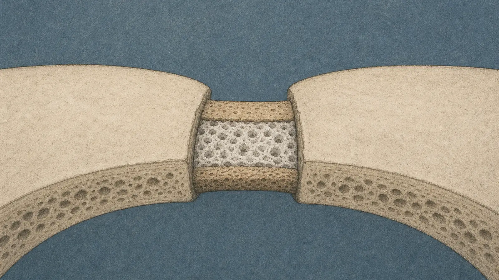
The operation is exactly as brutal as it sounds. A surgeon opens the scalp of an infant, sometimes only a few months old, lifts sections of the skull, and cuts out the bony seam that fused too early. That seam is a suture, and in a healthy baby it is not a defect but an engine: a thin band of unformed tissue where the skull keeps growing to keep pace with the brain underneath. In craniosynostosis, which shows up in roughly one birth in 2,500, one or more of those seams hardens shut too soon, and pressure can build inside a skull that no longer expands. The standard answer is to cut.
Here is the part the brochures skip. Sometimes the cut does not hold. The bone the surgeon just opened grows back together, the suture re-fuses, and the whole thing has to be done again. In a single-center review of 70 nonsyndromic cases, 6 children, about 8.5 percent, went back under the knife, one of them twice, because the seam closed again. Roughly one in twelve. In the harder syndromic anatomy the rate runs higher. The surgery, in other words, treats the shape of the problem without restoring the thing that was actually lost.
What was lost is a population of cells. In 2021, researchers established that skeletal stem and progenitor cells living inside the suture are what keep it open; thin them out and the suture fuses, which is craniosynostosis in miniature. That reframes the disease as a niche problem rather than a bone problem. You can shave and reshape all the bone you like, but if the stem-cell niche that maintains the gap is gone, the body will keep doing what bodies do, which is heal a wound closed.
So the trick is not to cut better. It is to put the niche back. A team led by Yuji Mishina at the University of Michigan School of Dentistry and W. Benton Swanson, now at Harvard’s dental school, built a scaffold to do exactly that, and the design has a deliberately unglamorous logic: bone, suture, bone. Published May 28 in Bone Research, it is made from poly(L-lactic acid), an FDA-approved biodegradable polymer, and printed in three connected compartments of different pore sizes. The outer two have geometry that coaxes the surrounding tissue to lay down bone. The central one does the harder job, tuned to keep skeletal stem cells undifferentiated, holding the gap open, refusing to ossify.
That resistance to ossification is the whole invention. In the lab, the central compartment kept its cells stem-like and stayed unfused even when the researchers flooded it with bone morphogenetic protein, the signaling molecule that normally drives those cells straight to bone. A structure that holds its ground under a hard push to fuse is precisely what the failed surgeries lack.
Then they tested it where it counts next, if not yet where it counts most. In a mouse model of midline craniosynostosis, chosen because it resembles the most common nonsyndromic human form, the team surgically removed the fused suture and implanted the scaffold. The treated animals kept an open, suture-like band of tissue and grew better across the face and skull. The controls re-fused, the way operated sutures so often do. “Our goal was not simply to reopen a fused suture,” Mishina said, “but to regenerate the biological niche that allows the skull to grow normally.” One detail is worth holding onto for the clinic that does not yet exist: the earlier they intervened, the better it worked.
Now the discipline, because it matters here more than usual. This is a mouse. The suture did not fail on its own; it was cut out by the same experimenters who then dropped their own device into the fresh defect, which is a long way from a squirming human infant with a syndromic skull and a surgeon watching the clock. And a scaffold that holds a gap open for the length of a mouse experiment is not the same animal as one that has to grow with a child for eighteen years, through every spurt the skull takes. The relevant question is not whether the tissue stayed open for weeks in a mouse. It is whether a biodegradable polymer can hand the job back to the child’s own biology before it dissolves, and that question the study cannot answer.
It sits on a ladder the same lab has been climbing, including earlier in vivo work on small-pore polymer scaffolds built to seed an artificial cranial niche. The Bone Research paper is the next rung, not the top. “Restores skull growth” is, for now, a sentence about mice. What the work actually offers is a mechanism-first alternative to a surgery whose signature weakness is that the body undoes it: the human skull is not a shape to be corrected once but a structure with its own architecture and growth math, and the cells that run that math are the thing this scaffold is trying to restore. That is a better question than the one the operating room has been answering for decades.
Whether the answer survives contact with a human trial is the whole matter from here, and that trial has not been run. For now the most honest sentence about the work is also the quietest one: in a mouse, the seam that was supposed to close stayed open, and the untreated ones next to it did not.
Sources
- News-Medical – Engineered scaffold restores skull growth in craniosynostosis mouse models (2026)
- Bone Research – Swanson, Mishina et al., multicompartment scaffold to regenerate the cranial suture stem-cell niche (2026)
- Nature Communications – Skeletal stem and progenitor cells maintain cranial suture patency and prevent craniosynostosis (2021)
- Turkish Journal of Medical Sciences – Amount of reoperation following surgical repair of nonsyndromic craniosynostosis at a single center (2022)
- Bioengineering – Initial in vivo analyses of small-pore polymer scaffolds for creation of an artificial cranial stem-cell niche (2026)
- The Cleft Palate-Craniofacial Journal – Craniosynostosis: Quantifying Differences in Skull Architecture (2025)