I used to think a T cell came with its job description printed at birth and kept it for life. Killer, helper, regulator: pick one, stick with it, die doing it. That model survived years of reading immunology. A paper out this week took it apart, and not in the way I expected. The donor cells that turn destructive don’t just misbehave. They change what they are depending on which layer of your gut they happen to be standing in.
On September 17, a team at Columbia published in Nature Immunology the most granular look yet at what a transplant recipient’s donor immune cells actually do inside the body, cell by cell, across two full years. They identified more than 51,000 alloreactive T-cell clones across 20 donor-recipient pairs, tracked 27 patients from day 3 after transplant out to day 732, and then went into the gut tissue itself with spatial sequencing fine enough to place a single cell in a single spot. The disease they were chasing is graft-versus-host disease, GVHD, and the cells tied to its worst form turn out not to be one fixed thing at all.
Here is the cruel geometry of a stem-cell transplant, the kind that saves people with leukemia. You wipe out the patient’s own marrow and blood system and replace it with a donor’s. The donor’s T cells come along for the ride, and those cells are useful: they hunt down any leukemia the chemo missed, the effect oncologists call graft-versus-leukemia. But the same cells can decide the entire patient is foreign and start attacking the gut, the skin, the liver. You cannot simply delete them, because the cells that cure and the cells that harm are cut from the same cloth. That is the knot GVHD has always presented, and it is why watching individual clones matters so much.
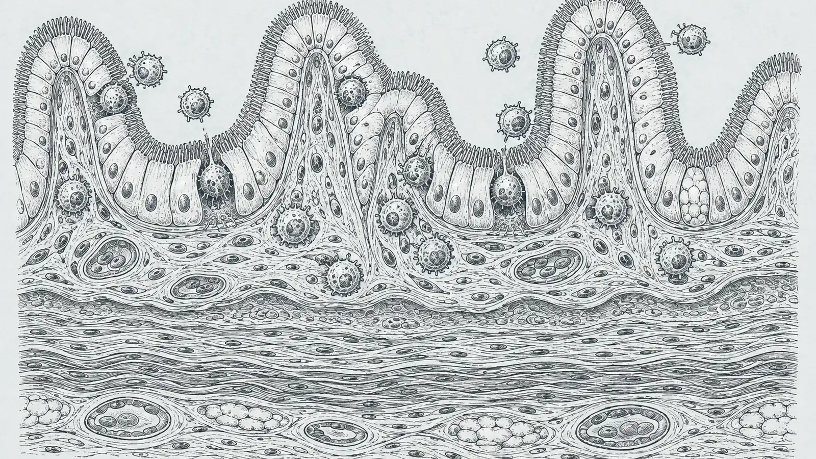
So what actually turns a helpful donor cell into a tissue-destroying one? This is where I had to stop and reread. Inside the gut wall there are two neighborhoods: the lamina propria, the loose connective layer underneath, and the intraepithelial compartment, the thin front line pressed right up against the cells that line your intestine. The Columbia team found the same CD8 killer clones sitting in both. And when a clone crossed from the lamina propria into that intraepithelial front line, it rewrote itself, switching on a tissue-resident memory program and becoming a “Hobit-positive” resident cell that no longer circulates and no longer leaves. The phenotype shift between the two layers was strong in GVHD patients (p < 0.001) and essentially absent in healthy donor tissue (p = 0.98). Wait, why would moving one cell-width over change what a cell is? Because the microenvironment does the reprogramming in place. The tissue talks to the cell, and the cell answers by becoming a permanent resident of the exact place it is damaging.
And the resident it becomes is a strange, contradictory creature. These Hobit-positive cells carry every exhaustion marker you would expect from a T cell that has been fighting too long, PD-1, CTLA4, TIM-3, the molecules that usually mean a cell is spent. Except they are not spent. They keep the cytotoxic granzymes GZMA and GZMB loaded, so they go on chewing at the epithelium while looking, on paper, half-dead. And when the team mapped where they park, the CD8 killers piled up in the exact regions where the gut’s regenerative crypts had been destroyed (p < 0.0001), nose to nose with the intestinal stem cells whose job is to rebuild the lining. They sit on the repair crew and keep it from working.
All of this leaves a signal in the blood long before a patient feels sick. Patients whose cumulative alloreactive-clone frequency stayed below 0.001 simply did not develop GVHD. Cross above 0.01 and you were looking at acute or chronic disease, most of it severe. The clones expand, quietly, in the weeks before the gut cramps and the diarrhea and the skin rash arrive. If you were counting, you would see it coming.
There is even a hint at why the standard rescue sometimes fails. In one severely affected patient, roughly 36 percent of a class of unconventional T cells had turned down NR3C1, the gene for the glucocorticoid receptor that steroids need to grab onto to work. Turn that receptor down and, at least on the transcriptome, the cell offers the front-line drug less to hold. It is a hint at steroid resistance in a single patient, not a mechanism anyone has tested functionally yet, and I want to keep it that size.
None of this means we have a therapy, and I want to be honest about the distance. The authors float their Hobit-positive residents as “an intermediate exhaustion state amenable to therapeutic intervention,” which is a hypothesis dressed as a finding. The obvious move, checkpoint blockade to un-exhaust or re-target these cells, is exactly the kind of thing that could make an autoimmune attack worse, and no trial has tested it here. This is 27 patients. The tissue biopsies came only from people who were already sick, so there is no matched healthy-gut comparison from the same patients, and the clone tracking read only the TCR beta chain, half of the receptor’s fingerprint. It is a map, a beautiful one, not a treatment.
But maps are how you find levers, and two of the levers here are not drugs at all. Post-transplant cyclophosphamide, a chemo given after the graft, pruned the alloreactive clones, cutting their frequency, number, and diversity (p < 0.0006) while barely touching the non-alloreactive ones, though it only worked if those clones had expanded early enough to be caught. And the gut’s own bacteria are in the loop: a separate 2024 mechanistic study in Immunity found the microbiota help dictate which T-cell clones get selected to expand and worsen GVHD in the first place. The organ where the war is fought and the microbes that live there are not bystanders. They are shaping who shows up to fight. That builds on earlier work in Science Translational Medicine showing donor CD8 cells adopt this tissue-resident, cytotoxic gut program early and that the signature tracks with the most severe grades of disease.
What I keep turning over is the timing. The danger is legible in a blood draw, in a clone count, weeks before anyone would think to intervene. Standard care still waits for the rash and the cramps and the diarrhea to announce themselves. If I were the one being tracked, I would want that clone count on my chart, and I would not accept watch-and-wait for symptoms as the whole plan.
Sources
- Nature Immunology – Shi et al., “Spatiotemporal single-cell profiling reveals T cell clonal dynamics and phenotypic plasticity in human graft-versus-host disease” (2026)
- PMC (open-access full text) – Shi et al., spatiotemporal single-cell analysis of T cell dynamics in human GVHD
- Science Translational Medicine – invasive and tissue-resident memory donor CD8+ T cells drive gastrointestinal acute GVHD (2021)
- Immunity – microbiota dictate T cell clonal selection to augment graft-versus-host disease after stem cell transplantation (2024)