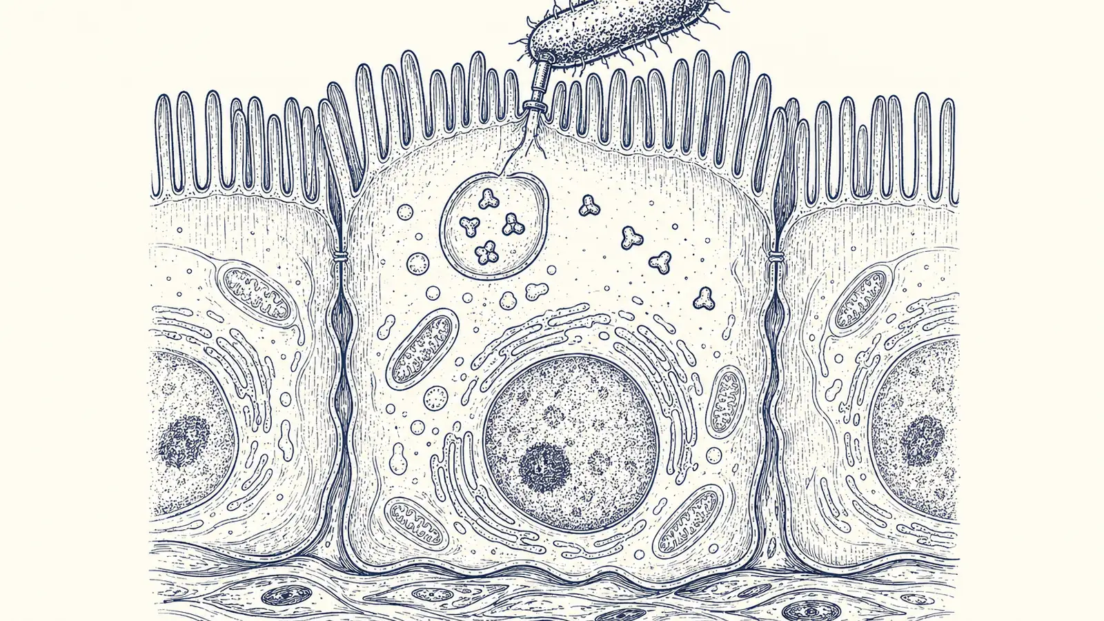
For as long as I have been writing about the gut, I filed the lining under plumbing. One sheet of cells, a single layer thick, doing the unglamorous work of letting nutrients in and keeping bacteria out. The immune teaching, I assumed, happened backstage, in the dendritic cells and lymph nodes that get paid for that job. Same flag, different address is the phrase I cannot stop repeating since reading a study published on September 30 in Nature Immunology, because the wall turns out to be a teacher, and it keeps its own files for months.
A team led by Casey Weaver at the University of Alabama at Birmingham reported that the epithelial cells lining the mouse colon present bacterial antigens directly to CD4 T cells, using MHC class II, the molecular display case immunologists have long treated as the business of professional immune cells. That display is what parks durable memory T cells inside the gut lining, where they sit long after the infection is gone.
The setup is clean enough that I want to walk you through it. The researchers used Citrobacter rodentium, the mouse stand-in for the strains of E. coli that give people food poisoning, and they rigged the bug to carry a single flag the immune system could track. Then they moved the flag around: stuck on the bacterium’s surface, pinned to the gut cell’s outer membrane, or injected straight through the bacterium’s type III secretion system, a literal molecular syringe, into the cytoplasm of the gut cell itself.
Only the flag delivered inside the cell produced much of anything. Injection into the cytosol drew 28 to 47 times more responding T cells than leaving it on the bacterial surface, and in the cleanest head-to-head, a full-length protein that gets injected versus a near-identical version that stays stuck to the bug, present in equal amounts, the injected one drove more than a hundredfold more memory cells. The authors put it flatly: “compartmentalization, not abundance, was the more critical determinant of immunogenicity.”
same antigen, delivered inside the cell
- Lower bound
- 28
- Upper bound
- 47
So why would the address of a molecule, rather than the sheer quantity of it, decide whether a gut bothers to remember a pathogen? Because of who can read it. The only cell positioned to see something floating in a gut cell’s cytoplasm, chew it up, and hand the fragment to a T cell is that gut cell. The barrier is not relaying the alert to someone more qualified. It is doing the presenting.
Then they took the display case away, and the lining’s role stopped being an inference. Deleting the epithelial MHC class II gene collapsed the pool of responding cells in gut tissue from about 1,317 to 53, more than a twentyfold drop. A labeling trick that tags cells which have physically touched an epithelial cell through MHC II showed 79 percent of the resident memory cells in the lining carrying that mark, and stripping the epithelial display case out dropped it to 16 percent. The wall was not incidental to the handshake. It was the handshake.
| Measure | Share |
|---|---|
| of resident memory cells | 79 percent |
The maintenance part is what rearranged my thinking. When the team deleted epithelial MHC class II after the infection had already cleared, the resident memory cells began to fade, so the gut cells have to keep presenting for the memory to stay parked. And stay it did: antigen-specific cells were still sitting in the lining at 240 days, eight months, long past the infection. Nor was it a faint trace. Primed mice hit later with a completely different bug, Salmonella, mounted more than ten times the T-cell response, poured out more of the inflammatory signal IFN-gamma, cleared the infection faster, and every one of them survived, while half the unprimed controls did not.
For decades the gut epithelium got written up as a passive barrier with a bit of immune chatter on the side, and the last few years have been dismantling that quietly. A 2023 paper in Nature had already shown that compartmentalized antigen presentation in the gut shapes immunity, and work in the lung found that epithelial cells there direct resident memory T cells as well. Your barriers are not bystanders in any of it. They are tissue that learns from what passes through, which makes the protective memory living at a surface look like something you grow by meeting the world there, through the ordinary traffic of a gut doing its job, rather than something delivered from outside.
The ceiling on all this is worth saying plainly, because the lean is earned rather than assumed. This is mice, and it is a single T-cell clone responding to one viral peptide used as a stand-in flag, which means the relevance to human E. coli is inferred and not demonstrated. Groups ran three to six animals, one imaging analysis rests on a single mouse, and the authors are refreshingly upfront that they do not yet know how an antigen sitting in the cytosol gets loaded onto MHC class II at all. They suspect autophagy, the cell’s recycling machinery, without having shown it. Early, taxpayer-funded basic biology from a university lab on NIH and NIAID money, not a clinical finding, and nobody should dress it up as one.
So here is where I have landed, and it is a change for me. I have stopped treating my gut lining as plumbing and started thinking of it the way I think of a lymph node, as tissue actively writing immune memory from whatever it meets. That shifts something small and practical. Nothing in this paper tested antibiotics, and I am not claiming it did, but if the lining is schooled by the microbial traffic crossing it, then wiping out that traffic is not a side effect I get to wave off: I will push harder next time a doctor offers me a course I do not clearly need.
Sources
- Nature Immunology (2026), Wilson, Acharya et al., open-access full text
- Nature Immunology, primary study (journal landing / DOI)
- Nature (2023), “Epithelial IFNγ signalling and compartmentalized antigen presentation orchestrate gut immunity”
- Nature Communications (2021), “Antigen presentation by lung epithelial cells directs CD4+ TRM cell function and regulates barrier immunity”