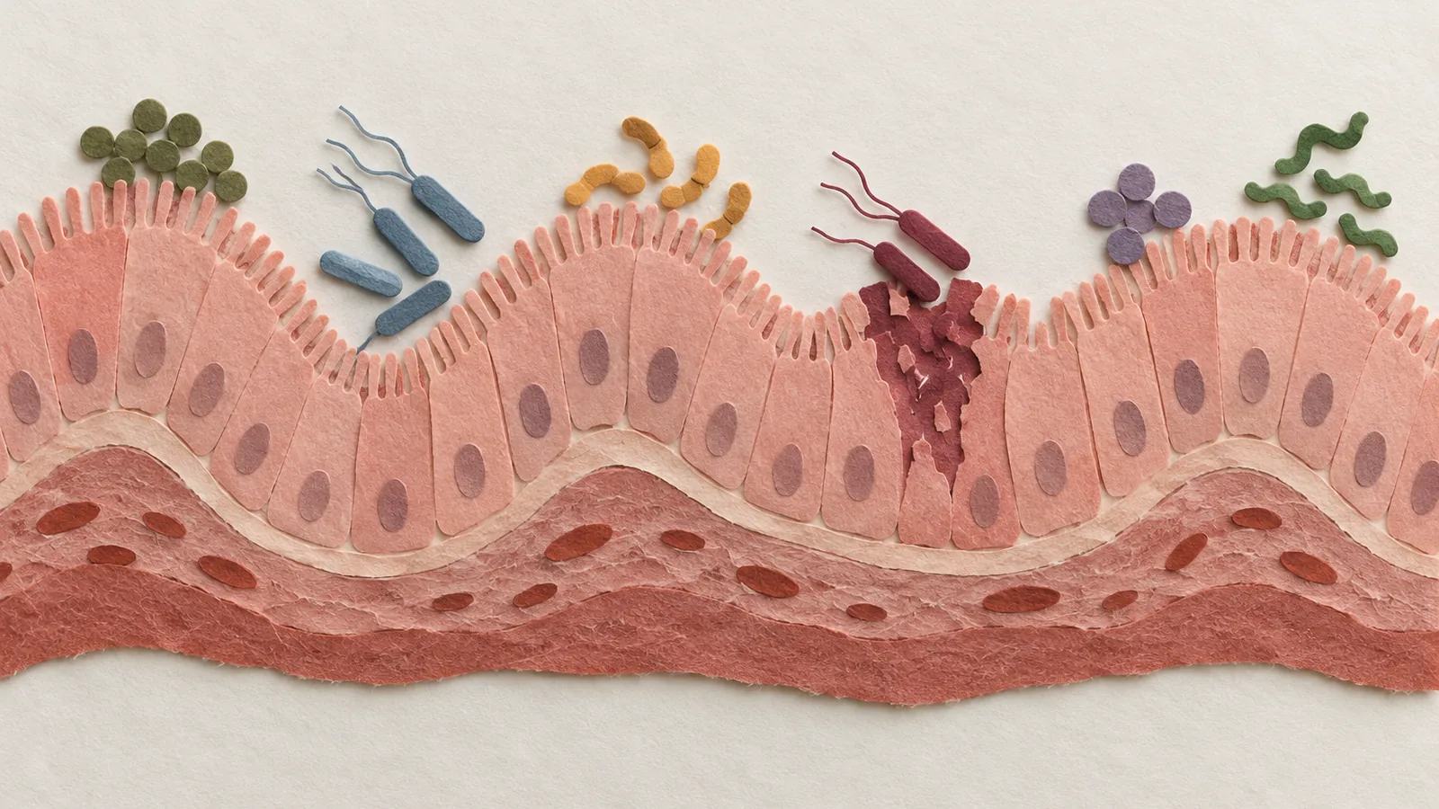
For years I filed the lining of my own gut under “plumbing.” One sheet of cells, a single layer thick, doing the unglamorous work of letting the good stuff in and keeping everything else out, while the real drama (the fighting, the deciding, the remembering) happened in the immune cells packed underneath it. That mental model just took a serious hit, and I am still turning over how wrong it was.
A team led by Daniel Beiting at the University of Pennsylvania and David Artis at Weill Cornell sat down and mapped 505,956 individual cells from the mouse small intestine and its draining lymph nodes, reading out both the genes each cell was running and the proteins on its surface, then sorting them into 91 distinct cell states in the ileum alone. They called the atlas GutPath and published it in Nature Immunology on October 7. Then they did the thing that makes it more than a pretty catalog: they infected the mice with six very different invaders and watched the whole sheet respond. A parasitic worm, the fungus Candida albicans, the parasite Cryptosporidium, a norovirus, and two bacteria that could hardly be less alike, the gut commensal known as segmented filamentous bacteria and the invasive pathogen Yersinia pseudotuberculosis.
You would expect the immune cells to light up, and they did. But when the team ranked which cells changed the most, the lining itself was out front. Enterocytes, the absorptive workhorses, along with gut stem cells, their fast-dividing daughters, goblet cells, and the fibroblasts beneath them, were the main drivers of the response across every single infection. The supposedly passive barrier was doing a lot of the talking.
And the specific genes those lining cells switched on barely overlapped from one infection to the next. Same cell types leading the charge, almost entirely different scripts. The worm’s enterocytes and the bacterium’s enterocytes were reacting to their own separate wars. So the gut lining is not running one generic “I am under attack” alarm. It is reading who the attacker is and answering in a dialect built for that specific enemy. How does a cell whose day job is absorbing amino acids know the difference between a tapeworm and a norovirus well enough to pick a custom response? That is the question the atlas opens and does not fully close.
It gets stranger with Yersinia, the invasive one. During that infection the team found an enterocyte state that simply does not exist in a healthy gut: a cell identity defined by 132 genes, including the acute-phase proteins Saa1 and Saa2 and the antimicrobial effectors Nos2 and Reg3a, with the transcription factors STAT3 and SREBF1 scoring as its likely conductors. A nutrient-absorbing cell had been repurposed into something closer to an inflammatory sentinel. And when they looked at where these cells physically sat, the signal peaked right on top of the pyogranulomas, the dense knots of inflammation that Yersinia builds and that are themselves packed with the bacteria.
Wait, why would an absorptive cell reinvent itself as a frontline fighter, and only inside the lesion where the bug is thickest? The researchers sketch a proposed wiring diagram: immune cells release the signal IL-22, that nudges STAT3 inside the enterocyte, and the enterocyte answers by pumping out IL-18, feeding the inflammatory loop right back. This is a hypothesis they float, not a mechanism they nailed down, and I want to be clear about that. But even as a hypothesis it moves the enterocyte from spectator to participant in its own defense, and it is exactly the kind of local, spatially pinned conversation you would never catch by grinding up a whole intestine and averaging it.
Then there is the part that sits squarely in my wheelhouse, which is what all this does to how the gut handles food. Of 134 metabolic and nutrient-transport genes the team tracked, 51 normally shift as an enterocyte matures, and 20 of those got rewired by at least one infection. The worm, Nippostrongylus, by the atlas’s metabolic read, dialed down the enterocytes’ secondary bile acid synthesis and ramped up fatty acid breakdown. Cryptosporidium and the colonizing bacteria both drove a loss of mature enterocytes. So the lining does not just fight; it reroutes its own metabolism in the middle of the fight, and it does so differently depending on who it is fighting. That lands for me, because it offers a plausible lead on something people describe all the time and clinicians struggle to explain: why two gut infections can leave you feeling metabolically different on the way out, long after the bug is gone. Whether that holds in a human gut is the next question, not a settled one.
| Group | Value (genes) |
|---|---|
| Tracked in enterocytes | 134 |
| Shift as the cell matures | 51 |
| Rewired by infection | 20 |
Now the honest fine print, and it cuts the other way from how fine print usually reads. This is mice, not people. It is a single early snapshot, under a week after each infection, so we do not yet know which of these programs stick around and which fade, and the authors say so plainly. A half-million-cell atlas is a map, not a clinical trial. None of that shrinks the reframe, though. It tells you the epithelium has been badly undersold, cast as the wall when it is closer to a combatant with a different battle plan for every enemy.
That reframe is where I will actually change something. I have spent a lot of words, in my own head and on this page, treating “leaky gut” and post-infection gut trouble as vague immune noise with the barrier as an innocent bystander. I am done doing that. The lining was never plumbing; it may be remodeled by the specific fight it just survived. So the next time I read about lingering symptoms after a gut infection, I am going to ask what the lining itself was rebuilt into, and whether it ever fully changed back, instead of assuming the barrier quietly reset once the fighting stopped.
Sources
- Nature Immunology – Beiting, Artis et al., “Diverse infections transcriptionally reprogram the intestinal epithelium and epithelial–immune cell interactions” (Oct 7, 2026)
- GutPath – open full text: 505,956 single cells, 91 ileal cell states, the Yersinia-specific enterocyte state, and the infection-specific nutrient-transport changes