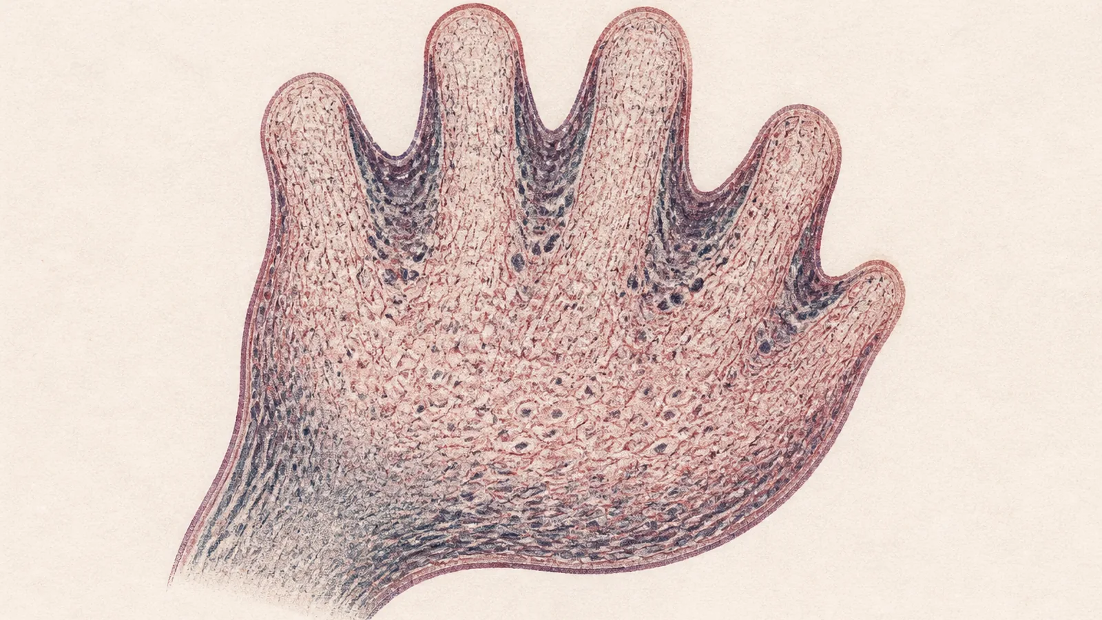
For years I filed cellular senescence in a mental folder marked damage. Senescent cells were the rust: worn-out, won’t-divide, won’t-die, piling up as you age, leaking inflammation into the tissue around them, turning up in the biology of arthritis, fibrosis, and frailty. Clear them out, the pitch goes, and you slow the decline. There is now a whole market built on exactly that premise, bottles and IV drips sold on the promise of fewer zombie cells. So the finding that stopped me cold this month is almost rude in how cleanly it flips the story: before any of us was old enough to leak inflammation into anything, we used the very same machinery to build a body. Senescence sculpted your fingers before it ever aged your skin.
The program we call "aging" was a construction tool first.
Nature Immunology ran a feature on embryonic senescence this month, and the science underneath it has been quietly dismantling the “senescence equals aging” assumption for more than a decade. Two 2013 papers in Cell, working in mouse and chick embryos, found senescent cells sitting in oddly precise locations, and not by accident. They cluster at the apical ectodermal ridge, the ribbon of tissue at the tip of a limb bud that tells fingers where and how to form. They show up in the endolymphatic sac as the inner ear takes shape, in the neural roof plate, and in the mesonephros, a throwaway embryonic kidney that has to be demolished on schedule. Same cell state we blame for getting old. Deployed, on purpose, to make a person.
So why would an embryo want cells that have stopped dividing? An embryo is supposed to be nothing but growth and division, and here it is deliberately switching cells into a state whose entire definition is they quit multiplying. Because a senescent cell does not just quit. It secretes. That much-maligned senescence-associated secretory phenotype, the SASP, the cocktail of signals we usually describe as inflammatory pollution, is in the embryo an instruction. The cell sits in one spot, pours out signals that shape how the neighboring tissue grows and folds, then hands itself over to be eaten. And here is where the wiring diverges from the aging version: developmental senescence leans on p21 but runs independent of p53, a different circuit from the DNA-damage senescence of old tissue. Knock p21 out of a mouse embryo and you get real patterning defects. The program we call “aging” was a construction tool first.
Then there is the cleanup, and this is the part I keep turning over. The macrophages that arrive to digest these cells are not a janitorial crew showing up after an accident. In those 2013 developmental studies they are finishing a job the senescent cells started: the dying cells recruit the very immune cells that come to remove them, and the tissue gets remodeled and reset. Wait, why would a cell about to be eaten call in its own executioner? Because that is the intent. The immune system here is not surveillance against decay. It is the sculptor’s assistant, chiseling away the scaffold once the shape is set, a senescence-and-clearance dynamic that shows up again and again across normal physiology, not only in aging tissue.
The same tool in the wrong hands ruins the work. A 2025 study built blastoids, lab-grown stand-ins for the very early embryo, from totipotent cells, then hit those cells with a jolt of hydrogen peroxide to force senescence early, before the developmental program called for it. The senescence markers lit up, p16 and p21 and TNF-α climbing while the totipotency genes faded, and the structures fell apart in exactly the ways that matter for a pregnancy. Cells sorted into the wrong lineages. When transferred into mouse uteri, the blastoids implanted poorly and built far less of the decidual tissue a pregnancy depends on. The authors don’t claim to have shown this in women; they propose the model as a window onto human infertility and the falling pregnancy rates of advanced maternal age, which is the honest framing for a mouse-and-dish experiment. Senescence in the embryo is neither good nor bad. It is constructive when it lands in the right place at the right moment, and corrosive when it fires too early in the wrong cell. Timing decides which one you get.
That is the complication the longevity market would rather you skip. If senescence is a signaling state your body reaches for on purpose, then “clear out all the zombie cells” stops being the obvious win the marketing implies. Context decides whether a senescent cell is sculpting or rotting, and a capsule swallowed at breakfast does not know the difference. It gets worse for the tidy version. Even the workhorse lab stain used to find senescent cells, SA-β-galactosidase, is shaky enough in living tissue that a 2021 paper put the doubt right in its title, asking whether it is a reliable in vivo marker at all. We are being sold precision against a target we cannot reliably see.
None of this means the aging version is a fiction. Senescent cells really do accumulate in old joints and scarred lungs, and the case for eventually targeting them there is not nonsense. What’s nonsense is the retail leap from that biology to a bottle: taking a process your body uses to build fingers and clear kidneys and reframing it as pure decay you can buy your way out of. I have stopped treating “fewer senescent cells” as a synonym for “younger,” and I will not hand money to a supplement sold on that equation. When the same process is both the sculptor and the rust, what matters is dose and timing, and right now nobody selling me a longevity capsule can tell me either one.
Sources
- Nature Immunology – “Embryonic senescence” feature (June 2026)
- Storer et al., Cell (2013) – Senescence is a developmental mechanism that contributes to embryonic growth and patterning
- Muñoz-Espín et al., Cell (2013) – Programmed cell senescence during mammalian embryonic development
- Nature Reviews Molecular Cell Biology – Developmentally programmed senescence
- Journal of Advanced Research (2025) – Senescence, implantation, and early embryonic development in totipotent cell-derived blastoids
- Journal of Translational Medicine (2026) – Immunological consequences of senescence in physiology and pathology
- Frontiers in Cell and Developmental Biology (2021) – Is senescence-associated β-galactosidase a reliable in vivo marker of cellular senescence during embryonic development?