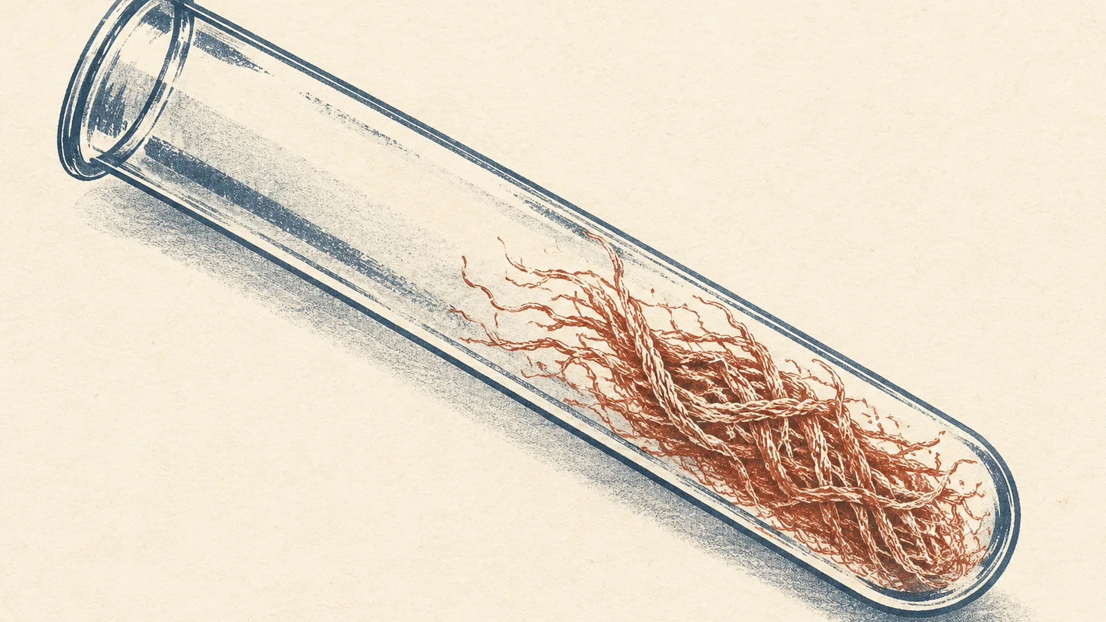
A fragment of the Alzheimer’s precursor protein that the field has called “non-amyloidogenic” for decades builds amyloid fibrils faster than amyloid beta does, at least in a test tube. I’ve read that phrase in so many papers that it had gone invisible to me, like the speed-limit sign on my own street, so my first reaction was a slightly embarrassing question: who tested the label before everyone started repeating it?
This is exactly the opposite of what we have actually shown
The fragment is called p3. Jevgenij Raskatov, a chemist at UC Santa Cruz, wants it renamed amyloid alpha, or Aα, and he makes the case in a ChemBioChem commentary titled “Amyloid Alpha: The Neglected Cousin of Amyloid Beta”, which ScienceDaily picked up this week under a “hidden culprit” headline. The commentary argues from published work rather than new experiments. “The P3 peptide is, most likely, not the innocent bystander it was commonly thought to be,” he told UC Santa Cruz’s news office earlier this year.
Two pairs of scissors, one leftover
The biology is simpler than the jargon. The amyloid precursor protein, APP, sits threaded through a neuron’s membrane like a pin pushed through fabric, and enzymes snip it. In the pathway everyone fears, beta-secretase cuts near the front of the amyloid beta sequence, gamma-secretase makes a second cut down inside the membrane, and out drops amyloid beta, usually the 40- or 42-residue version, with Aβ42 the stickier of the two.
Alpha-secretase cuts somewhere else, straight through the middle of the amyloid beta sequence between residues 16 and 17. Because that cut means full-length amyloid beta never forms, textbooks treat it as the moment the danger ends. But gamma-secretase still makes its snip, and what falls out is residues 17 through 40 or 42. That leftover is p3.
So why would anyone assume it was harmless? It keeps the back two-thirds of amyloid beta, and that’s where the clumping happens. In a 2020 review in the Journal of Alzheimer’s Disease, Raskatov and his then-graduate student Ariel Kuhn ran a standard aggregation-prediction tool and found that every predicted “hot spot” in amyloid beta sits inside the p3 stretch. The alpha cut throws away the front sixteen residues and leaves the sticky core intact. As they traced it, the label grew less from a decisive test than from a reputation: the alpha pathway was assumed to be protective, p3 was even floated as neuroprotective, and early electron microscopy described its deposits as amorphous rather than fibrous. Yet the warning was already in print. A 1995 study they cite found about 80 percent of p3 settling out of solution as aggregates, against about 40 percent of amyloid beta.
Faster builder, weaker poison
Raskatov says his lab has published three major papers over the past five years showing p3 forms amyloid at least as readily as amyloid beta. The most thorough independent check comes from John Viles’s group at Queen Mary University of London, whose February 2025 paper in Nature Communications put synthetic p3 through a full biophysical workup in test tubes and cultured cells.
Under matched conditions, p3 reached half-maximal aggregation in less than half the time full-length amyloid beta needed. Its growth was driven by secondary nucleation, meaning the surface of each finished fibril acts as a workbench where new fibrils get built, and under the microscope p3-40 and p3-42 fibrils carried roughly the same long and tight twists as Aβ40 and Aβ42. The Viles team wrote flatly that the p3 peptides “have been mis-named ‘non-amyloidogenic’.”
Then came the result that made me sit up. The fibrils seed each other. p3-40 seeds cut the lag time before Aβ40 started aggregating by almost half, and p3-42 had a similarly strong effect on Aβ42, but only when the tail ends matched. Wait, why would a fragment care which tail its cousin carries? Because the shared back end is the part that stacks into the fibril, so a matched p3 seed is effectively a ready-made template for amyloid beta to build on. The authors say this cross-seeding “may occur in vivo” but that proving it will take brain-derived p3 fibrils, which nobody has tested yet.
On toxicity, the honest reading is “less harmful, not harmless.” Amyloid beta oligomers let calcium pour into human cells in almost every preparation; p3-40 and p3-42 oligomers did it in 54 and 64 percent. p3 oligomers reduced cell viability, less than Aβ42 did. And p3 formed little doughnuts, rings about 7 to 10 nanometers across with a 1-to-2-nanometer hole through the middle, the shape you’d expect of a pore. p3-42 opened working ion channels in 16 percent of patch-clamp recordings, against 35 percent for Aβ42, a gap the authors called significant at P < 0.1 by a Fisher test, which is a looser bar than the usual 0.05.
| Group | Value (percent of recordings) |
|---|---|
| p3-42 oligomers | 16 |
| Aβ42 oligomers | 35 |
Under the same stain
The review sent me back to the pathology. p3 has been found in plaques from post-mortem Alzheimer’s brains, and Kuhn and Raskatov note it was identified as the major component of the diffuse plaques seen in Down syndrome; the Viles paper says it outnumbers full-length amyloid beta in cerebellar pre-amyloid deposits there. Then there’s the stain. 4G8, a widely used antibody for spotting amyloid in tissue, binds residues 17 to 24, which p3 and amyloid beta share. “The use of 4G8 would prevent any distinction between p3 and Aβ,” Kuhn and Raskatov wrote. That doesn’t tell anyone how much historical “amyloid beta” staining was really p3. It does mean the tool couldn’t have told them, and the review adds that standard extraction methods, built around amyloid beta’s chemistry, may not pull p3 out cleanly either.
The story also cuts the other way, and the “hidden culprit” headline skips it. Raskatov’s own lab published a 2024 Journal of the American Chemical Society paper, led by Kuhn, whose title says amyloid alpha “Attenuates Alzheimer’s Amyloid-β Toxicity via Cross-Chaperoning.” UC Santa Cruz’s summary says only that p3 may interact with amyloid beta “in ways that modulate its accumulation and toxicity.” A fragment that speeds amyloid beta’s assembly in one setting and blunts its toxicity in another is far more interesting than a villain, and far less settled.
A label that outran its evidence
None of that ambiguity rescues the old label, and some researchers have been reading the evidence backward. Raskatov told UC Santa Cruz he found at least four papers in peer-reviewed journals citing his lab’s work as proof that p3 is innocuous and does not form amyloid. “This is exactly the opposite of what we have actually shown,” he said. “We remain in the dark on how this sort of grand confusion may have come about.” The release doesn’t name the papers, and I’d love to see the list, because a field that cites a finding as its opposite has stopped reading its own footnotes.
Nobody is alleging fraud here, which separates p3 from the amyloid field’s worst recent embarrassment: the 2006 Nature paper that made the oligomer Aβ*56 a leading suspect in memory loss, retracted in 2024 after figures showed “signs of excessive manipulation, including splicing, duplication and the use of an eraser tool.” The p3 problem is quieter, a label repeated until it hardened into fact, but both failures grew in the same soil, a field far better at citing itself than at going back to check.
The money followed the label
UC Santa Cruz counts more than 400 Alzheimer’s drug trials, most aimed at amyloid beta and most failures. The flagship win shows how thin winning has been. In Eisai and Biogen’s Clarity AD trial of lecanemab, 1,795 people were randomized, and the drug slowed decline by 0.45 points on the 18-point CDR-SB dementia scale over 18 months, which got marketed as “27% slowing.” Brain swelling (ARIA-E) hit 12.6 percent of treated patients against 1.7 percent on placebo, and microbleeds and related findings (ARIA-H) 17.3 percent against 9.0 percent.
p3 also undercuts a quieter therapeutic idea. Because the alpha cut prevents full-length amyloid beta from forming, boosting alpha-secretase has been pitched as protective, and the Viles team now warns that producing p3 instead of amyloid beta “may in fact be undesirable.” Kuhn and Raskatov floated a hypothesis about the failed beta-secretase inhibitors along the same lines: the two enzymes compete for APP, so blocking one pair of scissors may simply route more protein to the other. They asked for trials to “quantify the levels of all products of AβPP in the blood and CSF.” UCLA neurology professor David Teplow told UC Santa Cruz the reevaluation “has far-reaching consequences for both basic science and clinical research.”
None of this shows p3 causes Alzheimer’s; the functional work is synthetic peptide in dishes, and these papers offer no measurement linking p3 levels in living patients to dementia. What it shows is that a fragment sitting in plaques, building fibrils faster than amyloid beta and forming pores in membranes, spent decades filed as safe on the strength of a reputation. If anyone ever asks me, or someone I love, to join a trial of a drug that pushes APP down the alpha pathway, I’ll ask for the p3 measurements first, and I won’t sign without them.
Sources
- Tian, Viles et al., Nature Communications (2025)
- Kuhn & Raskatov, Journal of Alzheimer’s Disease (2020)
- Raskatov, “Amyloid Alpha: The Neglected Cousin of Amyloid Beta,” ChemBioChem (2026)
- Kuhn, Raskatov et al., Journal of the American Chemical Society (2024)
- UC Santa Cruz News, “Supposedly harmless peptide may be linked to Alzheimer’s disease” (2026)
- Eisai, lecanemab Phase 3 Clarity AD full results (2022)
- Nature retraction note, Lesné et al. 2006 Aβ*56 paper (2024)
- ScienceDaily, “Scientists may have overlooked a hidden Alzheimer’s culprit for decades” (2026)